Expansive Techniques
Expansive techniques are utilized in the treatment of upper jaw atrophy and in cases where there is a displacement of between 3mm and 5mm in width between the corticals.
The techniques we use are the following:
SAGITTAL CREST OSTEOTOMY or SPLIT CREST
The residual alveolar crest is expanded by means of a green wood fracture of the vestibular cortical. The expansion of the crest can be carried out with an oscillating bone saw, with piezoelectric instruments or manually with a flat dental chisel.
OSTEOTOMY by means of crest expansion
This technique makes use of cylindrical osteotomes that, inserted into a crest of reduced width, but suitable depth, allow for the exclusive expansion of the site that will receive the implant, without involving the whole crest and thus resulting in far less trauma for the patient.
DISTRACTION OSTEOGENISIS
This technique allows for the formation of new bone between two bone segments, which after having been separated by means of osteotomy are slowly and progressively moved apart thanks to a distraction instrument.
The technique consists of three phases:
- Osteotomy and application of the distraction instrument with a seven day latent period.
- Distraction phase; commencing from the seventh day after the operation the distraction instrument is activated and begins to progressively widen the space between the two segments of bone. The vertical lift that can be achieved from this process varies from between 10mm to 15mm.
- Stabilisation phase; once the distraction is complete, having reached the desired height, the instrument is kept in place for a further 3 months so that a complete maturation of the new bone callus can occur. Distraction osteogenisis allows for the complete regeneration of atrophied bone, in addition to the above soft tissues, with notable aesthetic advantages for the patient.
LIFTING OF THE MAXILLARY SINUS
The maxillary sinus is a large cavity in the upper jaw, is connected to the nasal passages, is of pyramid shape and with age tends to grow in volume, meaning that, especially after the loss of premolars and molars, there is little bone in this region at the disposable of the oral surgeon for the insertion of implants and the re-establishment of chewing functions.
The aim of this treatment is to partially fill this cavity either with biomaterial mixed with PRP (agent of bone growth) or with autologous (from the patient) bone, thus obtaining at the end of the healing process a quantity of bone that is sufficient for implant insertion.
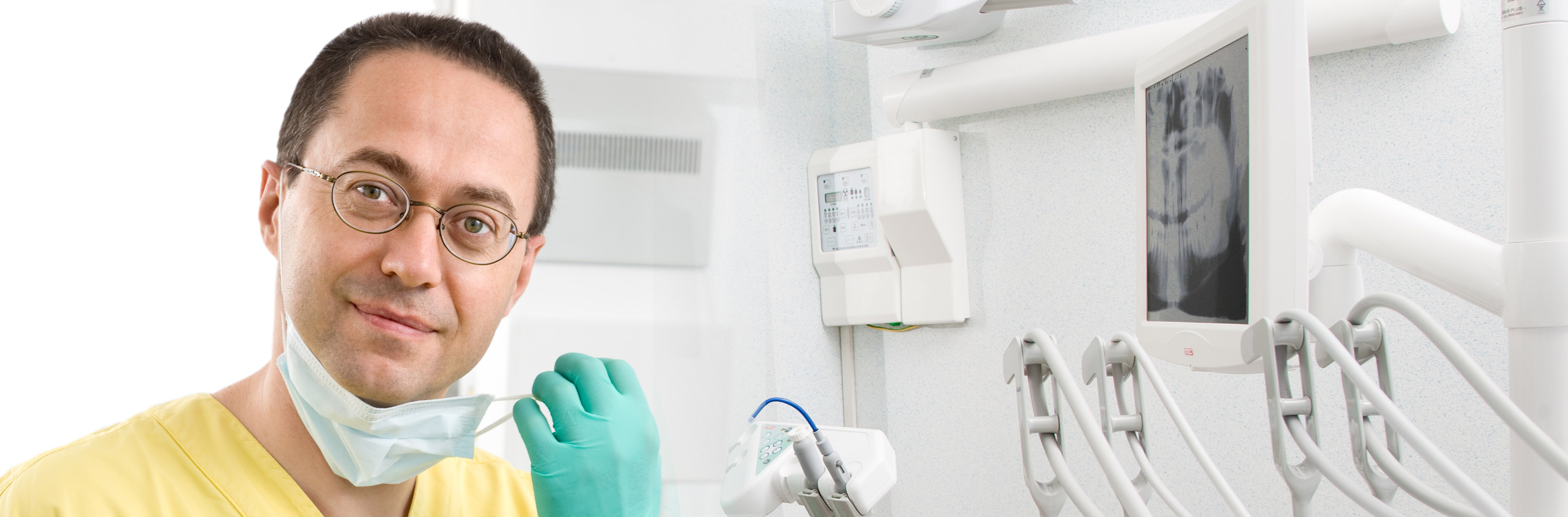
The pre-surgical radiography exams are crucial in defining the degree of bone atrophy around the jaw, both in the “reading” of the sinus’ anatomy and in the subsequent planning of the treatment.
The operations for the lifting of the sinus are of two types:
- Small sinus lift
Performed through the crest with osteotomes that allow for the lifting of the membrane which covers the internal side of the sinus and the subsequent insertion of either grafting or implantation materials. Also for this treatment we prefer to include PRP in the mixture of grafting materials.
- Large maxillary sinus lift
This technique consists of forming a window in the bone of the lateral sidewall of the sinus; the schneiderian membrane is peeled back and in the resulting space the filling material is inserted (here as well PRP is used though with significantly more need).